 Wohl Institute
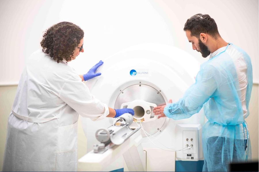
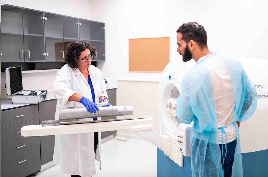


Preparation room
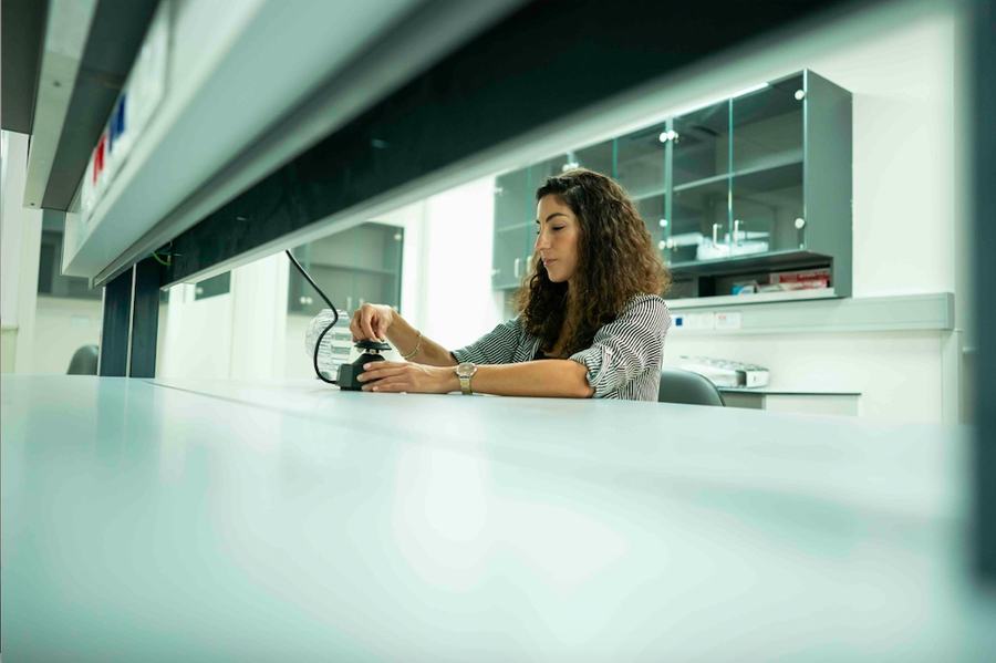
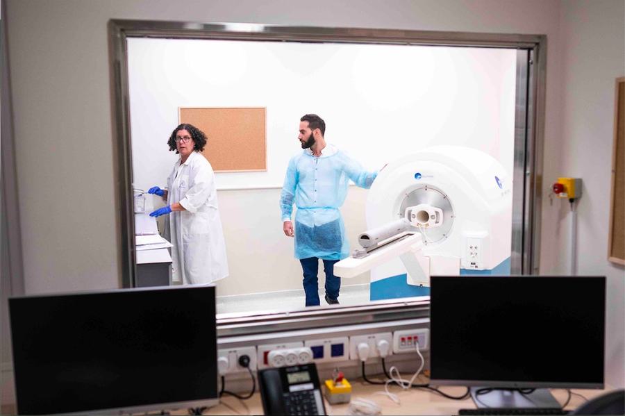
The facility possess a vast area to prepare the animals for the imaging; surgery, injection, shaving etc..
State-of-the-art imaging to follow disease progression in animal models, from molecular resolution up to in-vivo rodent studies.


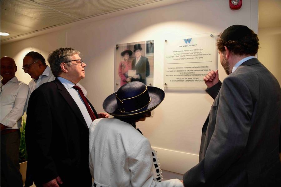
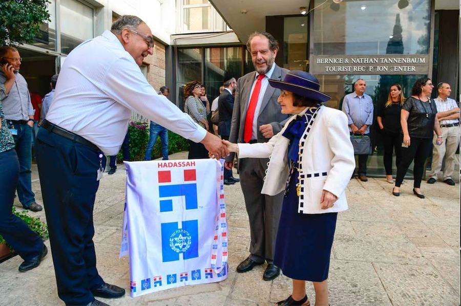
The Wohl Institute for Translational Medicine at Hadassah was established with a generous endowment from The Wohl Legacy Foundation. The Institute will advance the adaptation of clinical challenges into basic science laboratories to create disease models and develop novel therapeutic strategies from bedside to bench-top and vice versa.
The Wohl Institute offers state of the art imaging infrastructure enabling visualization and monitoring of disease progression using animal models.




Director of the Wohl Institute for Translational Medicine
Holds a PhD from the Weizmann institute and has 30 years' experience in preclinical advanced imaging modalities with emphasis in MRI technique development. During her twenty years employment at Hadassah she instructed numerous students on how to develop and utilize advanced imaging techniques.
The Wohl Institute, headed by Prof. Rinat Abramovitch, offers state-of-the-art technologies enabling visualization, digitization, and image analysis spanning from molecular resolution up to in-vivo rodent imaging. We provide our users with comprehensive one-stop imaging services that include technology implementation instructions for image acquisition and analysis.
We support researchers in
Six systems for anatomical, functional, metabolic and optical imaging, plus controlled-oxygen chambers.

The PET/MRI is a 7T cryogen-free magnet with a wide bore magnet to enable mouse and rat imaging. It includes PET insert that is based on silicon photomultiplier photo sensor technology for simultaneous imaging. This system enables anatomical, functional and metabolic imaging with high spatial resolution.
View details
The Siemens PET/CT system is a nuclear medicine technique which combines, in a single gantry, a positron emission tomography (PET) scanner and an x-ray computed tomography scanner, to acquire sequential images from both devices in the same session, which are combined into a single superimposed image.
View details
The Vevo3100 Ultrasound system is combined with LAZER-X system which enables photoacoustic imaging option. It operates with ultra-high frequency electronics up to 70 MHz, with spatial resolution down to 30 μm, suitable for in-vivo small animal imaging.
View details
Two optical imaging systems were purchased from Perkin-Elmer: the IVIS® Spectrum and the IVIS Lumina LT system. These high-throughput 2D optical imaging system combines high-sensitivity bioluminescence and fluorescence for in-vivo optical imaging.
View details
The U-CT system by MILabs is high-speed (5 s total body mouse) and can scan with ultra-low X-ray radiation doses, down to less than 2 mGy whole-body. Four performance levels ensure a system configuration that can be adapted to the user’s needs. By default, both mice and rats can be imaged.
View details
The OxyCycler A84XOV is a research tool for scientists who do oxygen sensitive work. It makes complex oxygen profile control easy. Control oxygen profiles with multiple setpoints anywhere from 0.1-99.9% oxygen.
View details
Director of the Wohl Institute

Lab Manager / Imaging Operator

Administrative Manager
Everything an imaging study needs, from animal preparation to data analysis.
The facility possess a vast area to prepare the animals for the imaging; surgery, injection, shaving etc..

A seminar room is available for planning future experiments or discuss the results obtained.

We also possess a Work Station to analyze your data after acquisition imaging.
