 FDG distribution
PET images living systems by recording high-energy γ-rays emitted from within the subject. The source of the radiation comes from positron-emitting-bound biological molecules, such as 18F-FDG (fludeoxyglucose), which is injected into the test subject. As the radioisotopes decay, they emit positrons which annihilates with electrons found naturally in the body. This produces 2 γ-rays at ~180° apart, which are picked up by sensors on opposite ends of the PET machine. This allows individual emission events to be localized within the body, and the data set is reconstructed to produce images.
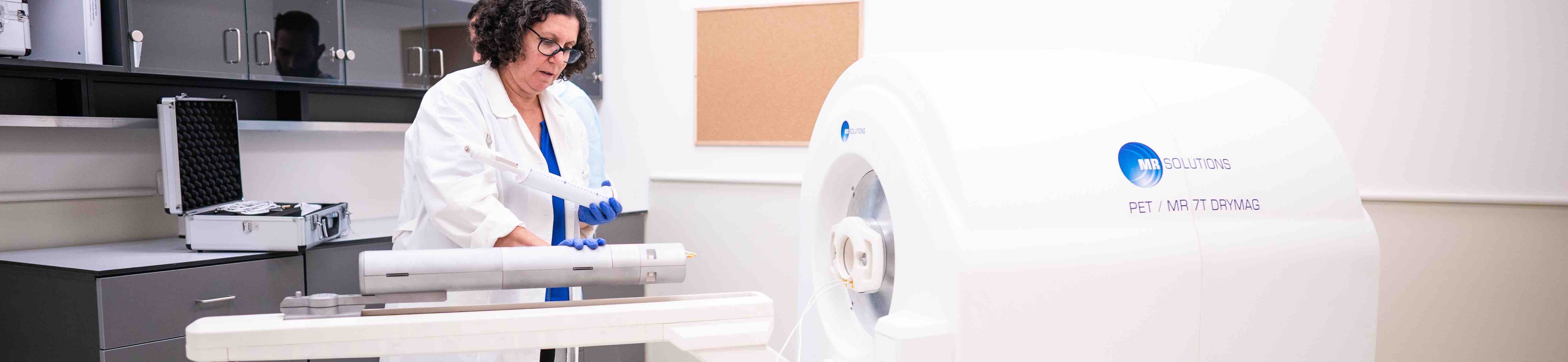
The MRS-7000 system allows simultaneous PET/MR imaging, and can image whole body mouse and rat heads.